A 2025 laboratory study found that oxalic acid can pull gadolinium from two MRI contrast agents and form a solid gadolinium compound. The result offers a possible explanation for some gadolinium deposits found after contrast-enhanced scans. It does not show that an ordinary meal before an MRI creates particles inside a person, or that changing diet prevents injury.
Gadolinium contrast can reveal findings that are hard to see on an unenhanced scan. Small amounts of the metal can also remain in tissues after the scan, which is why the chemistry deserves careful study. The study by Henderson and colleagues (https://pubmed.ncbi.nlm.nih.gov/40064247/) advances the chemistry while leaving the most important patient questions open.
What MRI contrast does
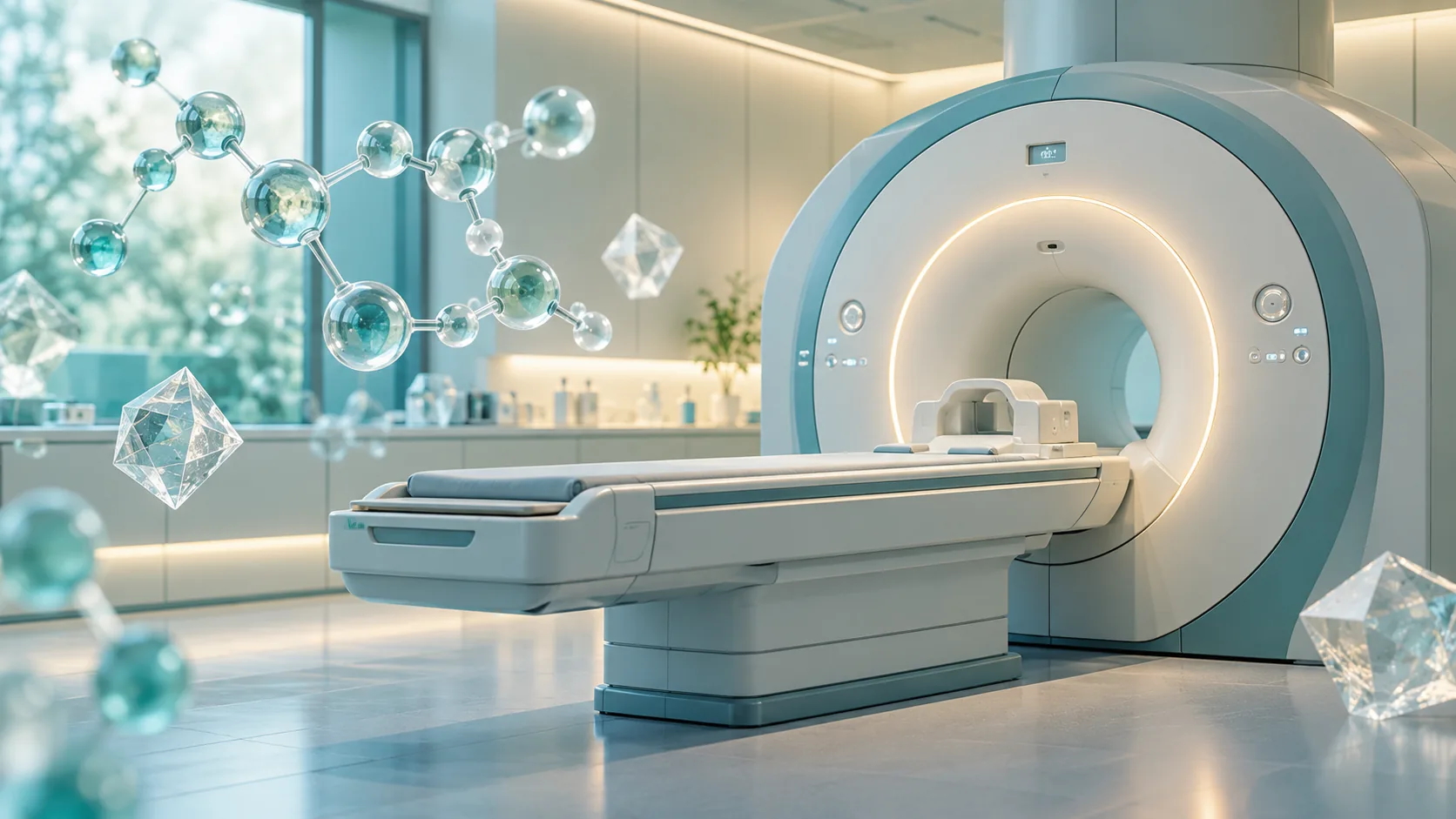
Some MRI scans use an injected gadolinium-based contrast agent. Gadolinium changes how nearby water appears on the image, which can make blood vessels, inflammation, tumors, and other findings easier to see. The metal is held by a carrier molecule, often called a chelator. The carrier is designed to keep gadolinium bound while the kidneys remove the agent.
Most of an injected dose leaves through the kidneys. Small amounts can remain in tissues for months or years, according to current US prescribing information for gadobutrol (https://dailymed.nlm.nih.gov/dailymed/drugInfo.cfm?setid=b7afe34d-b70f-41a5-b89c-561bdf29f046). Retention and harm are different findings. The label says the clinical consequences of retention have not been established in people with normal kidney function, while reports of symptoms after contrast exposure have not established a causal link. That statement does not mean every person's symptoms are understood. It means the evidence has not established the proposed link for that group.
Contrast products also differ. Some have a linear carrier, while others have a ring-shaped, or macrocyclic, carrier. Their stability and retention patterns differ. Those differences help clinicians choose an agent for a particular scan rather than treating every product as interchangeable.
The question the researchers tested
Researchers have previously identified gadolinium-rich particles in tissue after contrast exposure. Finding particles does not tell us exactly how they formed. The Henderson team asked whether oxalic acid, a compound made in the body and also related to some foods, could help separate gadolinium from its carrier and produce a solid deposit.
The team compared two marketed agents: Omniscan, a linear agent, and Dotarem, a macrocyclic agent. They mixed each with oxalic acid under controlled laboratory conditions. They then studied the solid product and timed how the Dotarem reaction changed with concentration, acidity, and added protein. This was a chemistry experiment, not a trial in patients. No group of people ate different foods before an MRI, and no clinical outcomes were measured.
The original paper (https://pubmed.ncbi.nlm.nih.gov/40064247/) reports that both tested agents yielded gadolinium oxalate. Omniscan reacted too quickly for the researchers to measure its reaction rate in the same way. Dotarem reacted more slowly, allowing them to examine which conditions promoted the visible precipitation. Even the ring-shaped agent formed a precipitate in the lab. The study did not measure how often, if ever, that reaction occurs after a clinical dose.
What changed the reaction
The Dotarem experiments revealed a strong dependence on the surroundings. In one series, the researchers held the Dotarem concentration at 166.7 millimoles per liter and varied the oxalic acid concentration from 33.3 to 333.3 millimoles per liter. Higher oxalic acid concentrations shortened the delay before a visible change and increased the measured precipitation rate. Changing the Dotarem concentration also changed the timing.
These concentrated mixtures made the reaction easier to observe. They do not describe the fluid around a patient's cells after an MRI. The paper does not show that normal blood oxalate reaches the same conditions or that a serving of spinach produces them.
Acidity was another key factor. A main mixture started at a pH close to 1.3, far more acidic than blood. The researchers adjusted the mixture toward less acidic conditions. Without added protein, they did not observe precipitation above roughly pH 2.2 in that set of experiments. With bovine serum albumin, a common laboratory protein, precipitation occurred at higher pH values, including values near those associated with acidic cell compartments called lysosomes. At pH 4.37, a higher protein concentration was needed for a reaction in the conditions reported.
Tissues contain proteins, and cell compartments called lysosomes are acidic. The laboratory mixtures captured parts of that environment while leaving out much of the body. A patient's tissue contains competing molecules, moving fluids, cells, and elimination pathways that a tube cannot reproduce. Even the paper's authors say their conditions do not necessarily reflect what happens to contrast agents in the body. Their work identifies a plausible pathway to investigate, not a measured rate of particle formation in patients.
Were particles found inside patients in this study?
No. This experiment characterized a precipitate formed from contrast agents and oxalic acid outside the body. Other research has reported gadolinium-rich particles in exposed tissue, including a study of renal tissue (https://pubmed.ncbi.nlm.nih.gov/36739294/). The tissue findings give researchers a reason to test this chemical route in living systems. The particles found so far have not all been identified as gadolinium oxalate.
The word nanoparticle can also suggest an established clinical threat when the crucial question remains unresolved: whether a given deposit causes injury, marks a process that causes injury, or stays in tissue without producing a measurable effect. A chemical identification and a health outcome are separate results. The 2025 study did not compare exposed people with and without symptoms, measure organ damage, or follow anyone after a scan. It cannot estimate an individual's chance of illness.
Claims that these particles act like sharp metallic shards should be treated carefully. The study identifies a chemical precipitate, not a demonstrated cutting action in human organs. It also does not show that particles produced in its lab setup enter the brain. Research on tissue retention remains important, but the mechanism of injury cannot be filled in with imagery.
The known serious risk: severe kidney disease
Gadolinium contrast has a well-established safety concern for people whose kidneys cannot clear it normally. Nephrogenic systemic fibrosis, or NSF, is a rare but serious condition involving hardening of skin and sometimes other tissues. Its risk has been strongly associated with certain contrast exposures in people with severe kidney dysfunction or acute kidney injury. Product labeling for gadobutrol describes this risk and calls for careful assessment of kidney function in relevant patients (https://dailymed.nlm.nih.gov/dailymed/drugInfo.cfm?setid=b7afe34d-b70f-41a5-b89c-561bdf29f046).
The oxalate experiment does not prove that gadolinium oxalate causes NSF. It also does not change the main clinical way this risk is managed: identify kidney problems, select an appropriate agent, and use contrast when the expected diagnostic benefit warrants it. People with known kidney disease, a recent kidney injury, or dialysis should tell the imaging team before a contrast scan. They should not assume an old kidney test represents their current condition if their health has changed.
For people with normal kidney function, gadolinium retention is documented, but the health consequences remain uncertain. Allergic reactions and other immediate side effects are separate concerns that a radiology team also considers. A clear discussion of benefit and risk should take account of the scan's purpose, the particular contrast agent, kidney status, previous reactions, and how many contrast scans a person may need over time.
Should you avoid spinach, chocolate, or vitamin C before an MRI?
The study gives no clinical basis for a pre-scan low-oxalate diet. Oxalate exists in the body, and foods can affect oxalate exposure, but the researchers did not test food intake, supplements, blood oxalate levels, or MRI outcomes in people. A concentrated oxalic acid reaction in a lab cannot be translated into instructions to avoid spinach, nuts, chocolate, or vitamin C for a specific number of hours or days.
A person with kidney stones or another condition may already have individual advice about oxalate or vitamin C. The new contrast study is not a reason to start a restrictive diet or stop prescribed supplements without discussing it with a clinician. It did not measure whether changing either one alters gadolinium retention.
The most useful step before a scan is to ask what information contrast is expected to add. A noncontrast MRI may answer some questions; other questions need contrast for a reliable diagnosis. If contrast is recommended, ask which agent will be used and whether your kidney history or previous contrast reactions change the plan. If you have had multiple contrast MRIs, mention them. These questions help the clinical team make a decision based on your actual scan and health history.
What would stronger evidence look like?
Researchers need to determine whether this specific gadolinium oxalate compound forms in living tissue after normal doses, under what conditions, and how much accumulates. The next challenge is linking a well-characterized deposit to a health outcome rather than assuming that any retained metal is harmful. Studies would need appropriate comparison groups, documented contrast agents and doses, kidney measurements, symptom assessments, and enough follow-up to distinguish a new problem from an existing one.
Independent replication matters because the 2025 work tested only two agents in a simplified system. Many contrast products are used clinically. Different carriers, doses, and tissue environments may change the chemistry. Studies that measure oxalate and other metabolic factors in patients could test the diet hypothesis directly. Until then, a person's diet cannot be used as a proven predictor of contrast injury.
Researchers now have a specific reaction to look for in tissue. This laboratory study involved no patients and measured no illness. If you have a contrast MRI scheduled, discuss your kidney history and the purpose of contrast with the imaging team. Do not delay a needed scan or change your diet on the strength of this lab finding alone.
This finding is another example of promising laboratory evidence that needs human testing. Our BPC-157 evidence review explains the same gap between a mechanism and a proven benefit.